I came across an article/report published in Nature. September 2016.
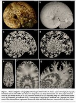
Suture pattern formation in ammonites and the unknown rear mantle structure
For some reason I had never thought of the ammonite as being fused to the shell. Fused is probably the wrong word. Honestly I had never thought about the specific anatomy and biology of ammonite or any of the shelled cephalopods with regards to their shells.
I think I must have had a vague notion that the relationship was somewhere between something similar to a clam with its shell (in terms of being attached, not how shell formation happens. Formation has no similarities whatsoever.) and a hermit crab with the shell it takes over. Now I’m Having a good laugh at myself for how I had never contemplated the anatomy and biology of ammonites.
I have frequently been curious as to how septa and sutures formed.
I absolutely love ammonite sutures. Sounds funny, but sutures have become a symbol of peace and serenity for me. It is a result of countless moments of sitting alone in some remote wilderness place, extracting an ammonite or baculites from the riverbed and sitting there in awe and wonder as I gaze at the beauty of the sutures. The water is babbling nearby and often the hues of sunset paint the horizon. So it’s a place of beautiful serenity. So when I look at sutures they have a calming affect and remind me of those peaceful, beautiful moments in the river. I digress.
I had never considered how the ammonite soft body parts may have looked. I had always assumed they looked like a squid. Therefore the concept of not being attached to their shell.
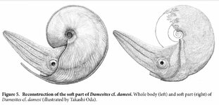
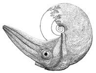
In this article is a drawing of the soft body parts of an ammonite. In the drawing the back end of the ammonite is depicted in the shape of its suture patterns. Much like how a sea cucumber cerata look. This makes a lot more sense than the body of a squid living in a shell. Each shelled mollusk, gastropods, bivalves, scaphopods, and cephalopods. . . each have a unique anatomy with relation to their shell and it’s formation that is quite different from the others.
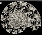
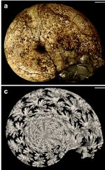
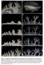
Reading this article really changed my visual concept of the anatomy of ammonites. Someone else may have posted this article before, but I thought I’d share it here for others who may not have read it. The images are beautiful. The CT 3D imaging is fantastic and so detailed.
These are images from the journal article.
Suture pattern formation in ammonites and the unknown rear mantle structure
For some reason I had never thought of the ammonite as being fused to the shell. Fused is probably the wrong word. Honestly I had never thought about the specific anatomy and biology of ammonite or any of the shelled cephalopods with regards to their shells.
I think I must have had a vague notion that the relationship was somewhere between something similar to a clam with its shell (in terms of being attached, not how shell formation happens. Formation has no similarities whatsoever.) and a hermit crab with the shell it takes over. Now I’m Having a good laugh at myself for how I had never contemplated the anatomy and biology of ammonites.
I have frequently been curious as to how septa and sutures formed.
I absolutely love ammonite sutures. Sounds funny, but sutures have become a symbol of peace and serenity for me. It is a result of countless moments of sitting alone in some remote wilderness place, extracting an ammonite or baculites from the riverbed and sitting there in awe and wonder as I gaze at the beauty of the sutures. The water is babbling nearby and often the hues of sunset paint the horizon. So it’s a place of beautiful serenity. So when I look at sutures they have a calming affect and remind me of those peaceful, beautiful moments in the river. I digress.
I had never considered how the ammonite soft body parts may have looked. I had always assumed they looked like a squid. Therefore the concept of not being attached to their shell.
In this article is a drawing of the soft body parts of an ammonite. In the drawing the back end of the ammonite is depicted in the shape of its suture patterns. Much like how a sea cucumber cerata look. This makes a lot more sense than the body of a squid living in a shell. Each shelled mollusk, gastropods, bivalves, scaphopods, and cephalopods. . . each have a unique anatomy with relation to their shell and it’s formation that is quite different from the others.
Reading this article really changed my visual concept of the anatomy of ammonites. Someone else may have posted this article before, but I thought I’d share it here for others who may not have read it. The images are beautiful. The CT 3D imaging is fantastic and so detailed.
These are images from the journal article.
Attachments
-
 7B7CFD25-022D-4EB2-818E-F97B8F706C03.jpeg300.5 KB · Views: 195
 -
 8F13791D-E659-4A6C-A41A-359A85E2961E.jpeg161 KB · Views: 185
 -
 F2E80BBF-DF3F-4BC1-BC40-94628B4571CA.jpeg365.3 KB · Views: 211
 -
 71FB1789-2664-4D94-B48B-4CADCDC48E79.jpeg227.1 KB · Views: 219
 -
 84F3654A-C099-4779-AD4E-982F6094A54F.jpeg222.9 KB · Views: 173
 -
 C0A93FDE-E72C-4C35-B4DA-0DA4A0B9DF57.jpeg520.8 KB · Views: 171
