[Octopus]: Iris - O. Briareus

SUPER photo (and nice crop pay off for taking a high resolution image)!!!! The chromatophores are really showing as well as the arms and eyes (edit: I guess you mentioned that in the post :oops:, can you tell I am excited?). I haven't located the dark spot for the hearts yet but I don't think I saw them at all for either of my tank hatches.
 
Last edited:
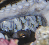
The latest look at the eggs. Iris has been really protecting (surrounding) them, so it's been a little more difficult to get a good view of the whole lot.
I'm not entirely sure what I'm looking at -- I had thought that the narrow dark part from the previous photos was the octopus body. But now it looks like there are chromatophores becoming visible just inside the egg casing. So does the body fill up the whole lower part of the egg? Is the darkest part part of the internal organs?
DSC_0003b.jpg

DSC_0003c.jpg


I got in my copepods, amphipods, mysids, and shore shrimp. The pods went into the buffet, and the shrimp into the main tank and the sump. Iris promptly snagged a few shrimp, so she's happy and well fed. Everything small is hard to see -- hopefully they're still around. The ghost shrimp are visible all over the place, but obviously too large for newborns to eat. Still very nervous about the hatch...
 
Those brown spots (you can see a few of them in prior photos) are not chromatophores but I am not sure what they are. I am thinking algae or reflection (possibly as an artifact of the flash). Try looking at a different time of day to see if they are still there and see if you can see them with your naked eyes (or try a non-flash picture). The body (mantle) is the long oblong shape behind the eyes, centered in the egg and about 1/2 the egg width (I think this is what you originally correctly identified as the body). The white blob in front of the eyes surrounded by the arms is the yolk sac and should be completely consumed when they hatch.

She may or may not be eating the shrimp. Her actions may be to keep the shrimp away from the eggs. Most brooding mothers stop eating completely but some have been known to eat a little during the brood. Those that do eat tend to live a bit longer but you will be surprised at what this has taken out of her when she emerges. The muscular arms will feel limp, her color will be quite gray and she will have difficulty controlling the stretched out mantle. Not every ending is the same but somewhat typically, she will stay in her den until her last day (or two) and then emerge to expire.
 
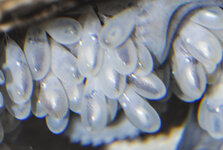
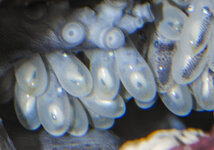
Some photos from this morning. I'm not sure that D's explanation of the spots on outer edge of the eggs is correct; I'm actually convinced that they are indeed chromatophores. Look at the following two photos, which were taken within a minute of each other. The camera didn't move, the camera settings were identical, and the photoshop "develop settings" were the same (essentially boosting both the brightness and contrast).

In addition to comparing the two photos directly, pay special attention (in the lower photo) to the pattern of dots compared to the position of the embryo within the egg. The embryos that are turned sideways (e.g. two of them on the top row in photo 2) also have a dot pattern that is turned sideways [not L/R symmetric]. Also, the embryos that are turned around backwards (e.g. the one at the lower right in photo 2) have a visibly different [but again L/R symmetric] dot pattern. Chromatophores!

On the other hand, I'm now confused again about what exactly I'm looking at -- namely, is the outer edge of the mantle right up against the egg casing, or is the dark central shape the mantle? I think, based on the chromatophores, the mantle must take up the whole lower volume of the egg, and that the dark shape is some other internal stuff. (Can you tell that I'm not a biologist?)
DSC_0005b.jpg

DSC_0006b.jpg
 
Glad you like it! I just borrowed a good macro lens from a neighbor, so the photos should improve a little (notice the incrementally improved focus on the last set).
 
Definitely not algae and 99% sure chromatophores. However, I think (note think, not know) it is reflective trickery. I can easily be mistaken and being sure would require the kind of examination only a few of us are willing to do (sacrifice an egg). I keep studying the photos (great shots!) and there are spots where they should not be (note the ones over the eyes). The animals are definitely inside a sac (review my empty eggs casings) so the outer shell is not their body but the part I can't swear to is where the sac ends and the body begins.

I can't be certain but I think you are getting flash reflection from the light bouncing inside the sac (notice how silver/shiny the parts of the body appear). This is a late stage photo of Kooah's eggs (the last in the group series that you can't enlarge), taken without flash (and sadly no macro lens). Note the chromatophores and the dark spots in the bodies that I believe are the primary hearts. Also look at the three top left eggs your post #30. You can clearly see chromatophores on the body but then the "floating" dots on the eggs.

eggs_20100525_cropped.jpg
 
Last edited:
Martin,
Never would have thought about "parts" but it seems reasonable and you are the only other member (welcome aboard!) who has had this success so ALL input is highly desirable! Of my brood, only 2 survived. Cross mating them produced hatchlings that died within a day or two (eggs started disappearing at an alarming rate about a week before hatching).
 
From today:
Really starting to see the form maturing!

The first photo is nice because it shows different embryos turned in many different directions; sort of a "survey" shot.

Note a couple of significant things in the second photo:
First, we're seeing the underside of the mantle in most of the eggs, and there is a dark, dare I say heart-shaped mark. What is this? Secondly, in the egg just right of center, you can barely make out the underside of the legs all spread out. It really looks like a little octopus for the first time!
DSC_0002b.jpg
DSC_0008b.jpg
 
Yes, the dark dots may be the branchial hearts (hearts for the gills). In the bottom photo, top layer, second octo, you can see both heart "dots" (I think :oops:). The sestemic (primary) heart is close by so in a small animal, it is harder to tell without seeing both sides. Here are a set of diagrams I found that labels the simplest internal components. I can't find much of a yolk sack (it will be a white ball grasped by the arms) so I think you are really close to seeing these little guys swimming about. It will likely happen at night so keep an eye out for "snow" in the tank that gravitates to the outer walls.
 
Last edited:
Thanks CG, I would not have considered that option but they do look below where the heart shadows should show. I have had dark spots on the mantel on the brain with three other, trying to decide if it is a species we don't usually see or if it is just the gill hearts oddly exposed through the skin.

@TMoct, I came across a list of linked external articles that used to be in the hatchling subsection that may be worth a look. It has been awhile since I have read them but it may help with activity while you twiddle your thumbs :wink:
 

Shop Amazon

Shop Amazon
Shop Amazon; support TONMO!
Shop Amazon
We are a participant in the Amazon Services LLC Associates Program, an affiliate program designed to provide a means for us to earn fees by linking to Amazon and affiliated sites.
Back
Top